BiologyUnit 167 min read
Developmental Biology: Fertilization to Embryo Formation
Unit 16 of Biology explores the fascinating journey from fertilization to embryo development, covering key stages like cleavage, gastrulation, and organogenesis, with clear diagrams and NEB-style questions to master the topic.
TAKEAWAYS:
- Developmental biology explains how a single fertilized egg (zygote) transforms into a complex multicellular organism through precise genetic and cellular processes.
- Key stages include fertilization, cleavage, gastrulation, and organogenesis, each with distinct cellular behaviors and outcomes.
- Induction and differentiation are critical mechanisms that guide cell specialization during embryogenesis.
- Model organisms like frogs and chickens help us understand human development due to conserved biological principles.
- NEB exams test your ability to label diagrams, compare stages, and explain processes—always use clear, step-by-step descriptions.
What is Developmental Biology?
Developmental biology is the study of how a single-celled zygote (fertilized egg) develops into a fully formed organism. This process involves cell division, cell movement, and cell specialization. Humans, like all animals, follow a predictable sequence of stages during embryogenesis. Understanding these stages helps explain birth defects, genetic disorders, and even how stem cells work.
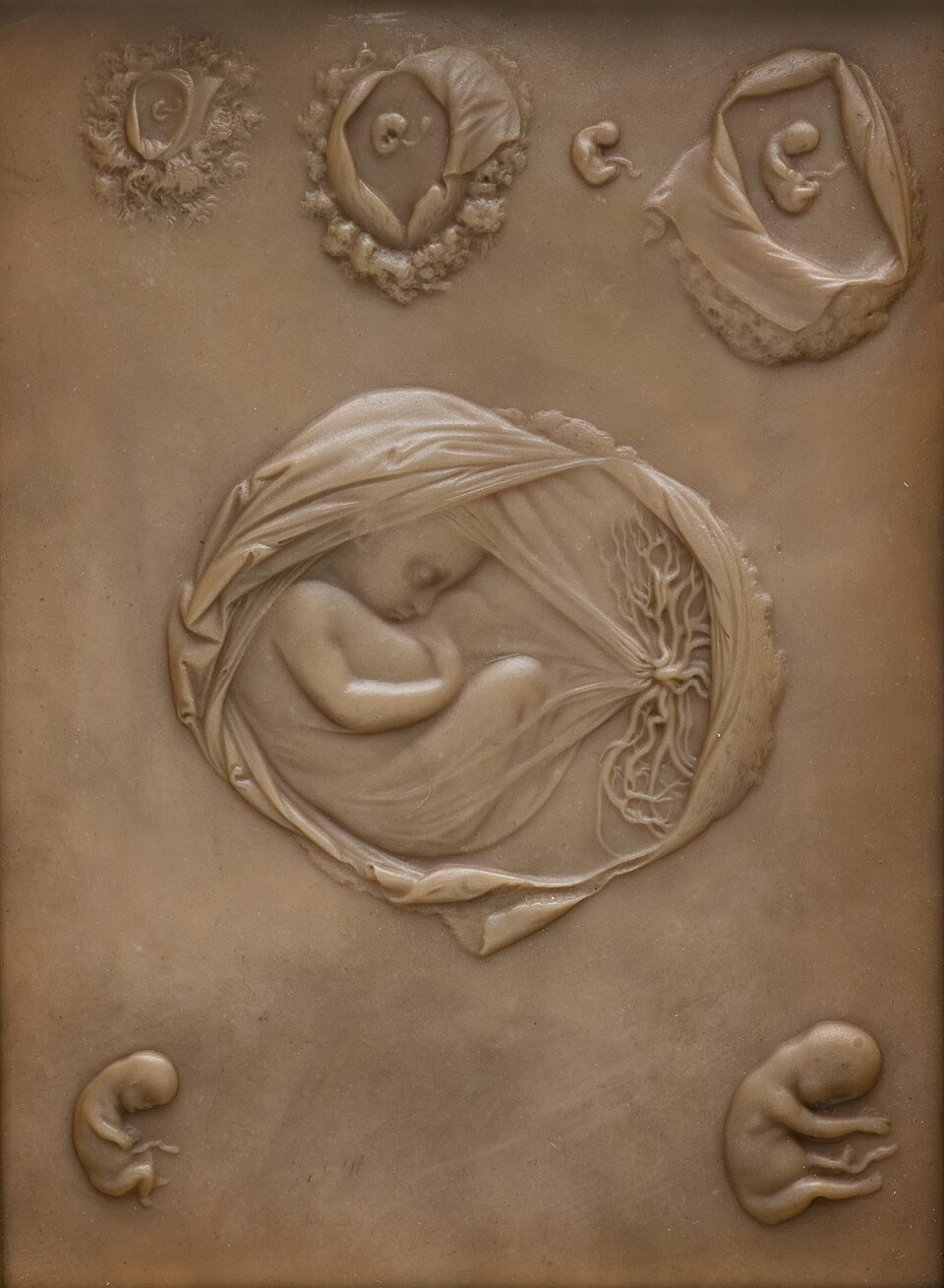 Key stages from zygote to fetus (weeks 1–8) (Image: CC BY 4.0, via Wikimedia Commons)
1. Fertilization: The Beginning of Life
Fertilization is the fusion of a male gamete (sperm) and a female gamete (egg/ovum) to form a zygote. This process occurs in the fallopian tube in humans and triggers the start of embryonic development.
How Fertilization Happens
- Sperm meets egg: Millions of sperm swim toward the egg, but only one successfully fertilizes it.
- Acrosomal reaction: The sperm releases enzymes to break through the egg’s outer layers.
- Fusion of membranes: The sperm’s nucleus enters the egg, and the egg’s membrane blocks other sperm (preventing polyspermy).
- Formation of zygote: The combined genetic material (23 chromosomes from sperm + 23 from egg) forms a diploid zygote.
flowchart TD
    A["Sperm + Egg"] --> B["Acrosomal Reaction\n(Enzymes break egg layers)"]
    B --> C["Fusion of Membranes\n(Only 1 sperm enters)"]
    C --> D["Zygote Formation\n(46 chromosomes)"]
    D --> E["Cleavage Begins"]
Importance of Fertilization
- Restores the diploid chromosome number (46 in humans).
- Activates the egg’s metabolism to begin development.
- Determines the genetic makeup of the organism.
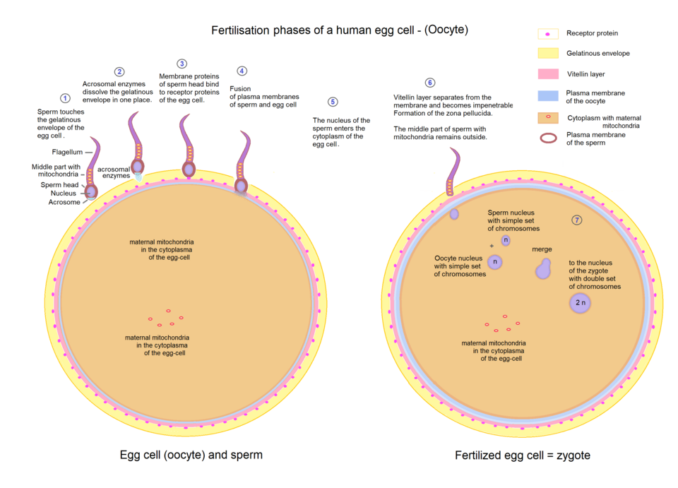 Label: Acrosome, egg membrane, sperm nucleus, zygote (Image: {{Sciencia58}}, CC0, via Wikimedia Commons)
2. Cleavage: Rapid Cell Division
After fertilization, the zygote undergoes cleavage—a series of rapid cell divisions without growth. This produces a multicellular embryo called a blastula.
Types of Cleavage
| Type | Description | Example Organisms |
|---|---|---|
| Holoblastic | Complete division of the egg | Frogs, humans |
| Meroblastic | Partial division (yolk-rich eggs) | Birds, reptiles |
| Rotational | Cleavage planes alternate directions | Mammals |
Stages of Cleavage in Humans
- 2-cell stage → 4-cell stage → 8-cell stage (first 3 days).
- Morula: A solid ball of cells (~16 cells).
- Blastula: A hollow ball with a fluid-filled cavity (blastocoel).
flowchart LR
    A["Zygote"] --> B["2-cell"] --> C["4-cell"] --> D["8-cell"]
    D --> E["Morula\n(Solid ball)"]
    E --> F["Blastula\n(Hollow ball)"]
3. Gastrulation: Formation of Germ Layers
Gastrulation transforms the blastula into a gastrula, which has three primary germ layers:
- Ectoderm → Nervous system, skin.
- Mesoderm → Muscles, bones, circulatory system.
- Endoderm → Digestive organs, lungs.
Process of Gastrulation
- Invagination: Cells move inward at the blastopore (opening of the gastrula).
- Formation of archenteron: The future digestive tract forms.
- Germ layer differentiation:
- Ectoderm: Outer layer.
- Mesoderm: Middle layer (forms between ecto- and endoderm).
- Endoderm: Inner layer.
flowchart TD
    A["Blastula"] --> B["Invagination\n(Cells move inward)"]
    B --> C["Gastrula\n(3 germ layers)"]
    C --> D["Ectoderm\n(Nervous system)"]
    C --> E["Mesoderm\n(Muscles, bones)"]
    C --> F["Endoderm\n(Organs)"]
4. Organogenesis: Formation of Organs
After gastrulation, organogenesis begins. Cells from the three germ layers differentiate into specific organs and tissues.
Key Processes in Organogenesis
| Germ Layer | Derivatives | Example Organs |
|---|---|---|
| Ectoderm | Nervous system, epidermis, lens of eye | Brain, spinal cord, skin |
| Mesoderm | Muscles, bones, heart, blood vessels | Heart, skeleton, kidneys |
| Endoderm | Digestive tract, lungs, liver, pancreas | Stomach, intestines, lungs |
5. Induction and Differentiation
- Induction: Signals from one group of cells tell others how to develop (e.g., notochord induces the neural tube).
- Differentiation: Cells become specialized (e.g., stem cells → nerve cells, muscle cells).
6. Model Organisms in Developmental Biology
Scientists study simpler organisms to understand human development:
- Frog (Xenopus): Easy to observe cleavage and gastrulation.
- Chicken: Meroblastic cleavage and organogenesis.
- Mouse: Similar to human embryology.
Exam Tip: How to Score Full Marks
- Label diagrams accurately: NEB often asks for labeled stages (e.g., morula, blastula, gastrula).
- Compare stages: Use tables to show differences (e.g., holoblastic vs. meroblastic cleavage).
- Explain processes step-by-step: Describe how and why each stage occurs.
- Relate to humans: Always connect model organisms (frogs, chickens) to human development.
- Use keywords: Terms like zygote, blastocoel, induction, differentiation fetch marks.
NEB-Style Questions & Solutions
Question 1 (Short Answer)
Describe the stages of cleavage in a human embryo.
Answer:
- Zygote → 2-cell stage (first division).
- 4-cell stage → 8-cell stage (rapid divisions).
- Morula (solid ball of ~16 cells).
- Blastula (hollow ball with blastocoel).
Marks: 1 mark per correct stage.
Question 2 (Diagram-Based)
Label the following parts in a gastrula: blastopore, archenteron, ectoderm, mesoderm, endoderm.
Answer:
graph TD
    A["Gastrula"] --> B["Blastopore\n(Opening)"]
    A --> C["Archenteron\n(Future gut)"]
    A --> D["Ectoderm\n(Outer layer)"]
    A --> E["Mesoderm\n(Middle layer)"]
A --> F["Endoderm\n(Inner layer)"]Marks: 1 mark per correct label.
Question 3 (Comparison)
Differentiate between holoblastic and meroblastic cleavage with examples.
| Feature | Holoblastic Cleavage | Meroblastic Cleavage |
|---|---|---|
| Definition | Complete division of the egg | Partial division (yolk-rich eggs) |
| Example | Frogs, humans | Birds, reptiles |
| Cleavage Type | Equal or unequal blastomeres | Disc-shaped cleavage (blastodisc) |
Marks: 2 marks for correct comparison.
Final Tip: Practice diagram labeling and stage descriptions daily. NEB loves detailed, step-by-step answers! 🚀
Based on the NEB +2 Science syllabus for Biology (Bio), unit 16.
Discussion
Loading…