BiologyUnit 1511 min read
Human Reproductive System: Anatomy, Physiology & Disorders
Unit 15 of Biology covers the male and female reproductive systems, gametogenesis, hormonal regulation, fertilization, pregnancy, and common reproductive disorders—essential for NEB exams with clear diagrams, flowcharts, and NEB-style questions.
TAKEAWAYS:
- The male and female reproductive systems have distinct organs and functions, but both rely on hormones (testosterone, estrogen, progesterone) to regulate development and reproduction.
- Gametogenesis (spermatogenesis in males, oogenesis in females) produces haploid gametes (sperm and egg) through meiosis, but sperm production is continuous while eggs are formed before birth.
- Fertilization occurs in the fallopian tube, forming a zygote that implants in the uterus after cleavage and blastocyst formation.
- Menstrual cycle (28 days) involves follicular phase (estrogen rise), ovulation (LH surge), and luteal phase (progesterone dominance), controlled by FSH and LH from the pituitary.
- STDs (e.g., HIV, syphilis, gonorrhea) and infertility (blocked fallopian tubes, low sperm count) are major disorders, often preventable with safe practices.
- Assisted reproductive technologies (IVF, IUI) help infertile couples, but ethical and social concerns exist.
1. Male Reproductive System
A. Anatomy
The male reproductive system produces sperm and hormones (testosterone). Key organs:
graph LR
    A["Testes"] -->|"Produces"| B["Sperm & Testosterone"]
    A --> C["Spermatogenesis"]
    B --> D["Epididymis"] -->|"Stores"| E["Vas Deferens"]
    E --> F["Urethra"] -->|"Exits via"| G["Penis"]
    H["Prostate Gland"] -->|"Secretes"| I["Alkaline Fluid"]
    J["Seminal Vesicles"] -->|"Adds"| K["Fructose & Prostaglandins"]
L["Bulbourethral Glands"] -->|"Lubricates"| G- Testes: Located in the scrotum (outside body for 3°C lower temp), produce sperm and testosterone.
- Epididymis: Stores and matures sperm (~2 weeks).
- Vas Deferens: Transports sperm to the ejaculatory duct.
- Seminal Vesicles: Add fructose (energy for sperm) and prostaglandins (stimulate uterine contractions).
- Prostate Gland: Secretes alkaline fluid to neutralize vaginal acidity.
- Bulbourethral Glands: Release pre-ejaculate to lubricate the urethra.
- Penis: Delivers sperm via urethra during ejaculation.
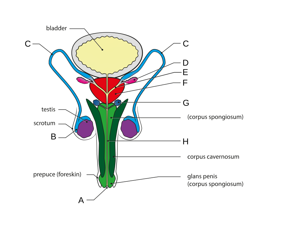 Label the testes, epididymis, vas deferens, urethra, and accessory glands. (Image: RWhitwam, CC BY-SA 4.0, via Wikimedia Commons)
B. Spermatogenesis
- Occurs in seminiferous tubules of testes.
- Stages:
- Spermatogonia (diploid) → Primary spermatocytes (meiosis I).
- Secondary spermatocytes → Spermatids (meiosis II).
- Spermatids → Spermatozoa (spermiogenesis).
- Hormonal Control:
- FSH (Follicle-Stimulating Hormone) → Stimulates sperm production.
- LH (Luteinizing Hormone) → Stimulates Leydig cells to produce testosterone.
- Testosterone → Supports sperm maturation and secondary sexual traits.
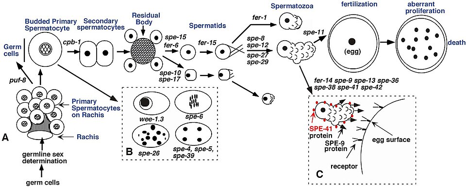 Show spermatogonia → sperm with labeled meiotic divisions. (Image: L'Hernault, S.W., CC BY 2.5, via Wikimedia Commons)
C. Semen and Ejaculation
- Semen = sperm (5%) + seminal fluid (95%) (from prostate and seminal vesicles).
- Ejaculation: Sympathetic nervous system triggers peristalsis in vas deferens → semen expelled via penis (~3–5 mL per ejaculate, 50–150 million sperm/mL).
2. Female Reproductive System
A. Anatomy
The female system produces eggs, supports fertilization, and nurtures a fetus. Key organs:
graph LR
    A["Ovaries"] -->|"Releases"| B["Egg (Ovum)"]
    A -->|"Secretes"| C["Estrogen & Progesterone"]
    B --> D["Fallopian Tube"] -->|"Fertilization site"| E["Uterus"]
    E -->|"Implantation"| F["Endometrium"]
    G["Cervix"] -->|"Connects"| H["Vagina"]
I["Vulva"] -->|"External parts"| J["Labia, Clitoris"]- Ovaries: Produce eggs (ova) and hormones (estrogen, progesterone).
- Fallopian Tubes: Site of fertilization; fimbriae sweep the egg into the tube.
- Uterus: Muscular organ where fertilized egg implants and fetus develops.
- Endometrium: Inner uterine lining, shed during menstruation.
- Cervix: Connects uterus to vagina; dilates during childbirth.
- Vagina: Birth canal and copulation organ.
- Vulva: External parts (labia, clitoris).
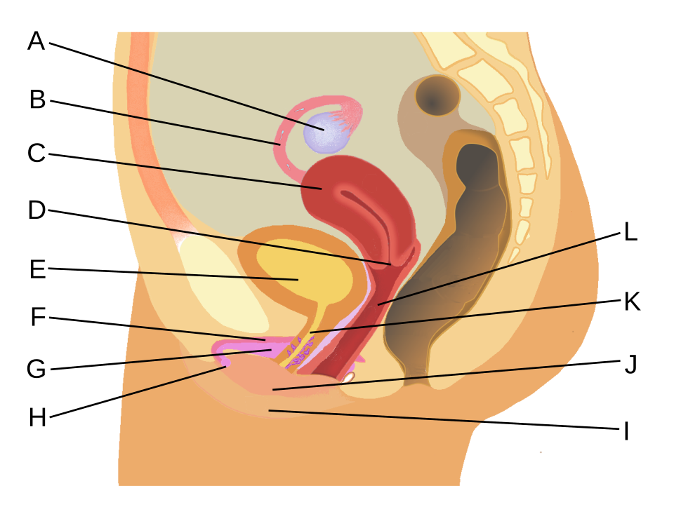 Label ovaries, fallopian tubes, uterus, cervix, and vagina. (Image: RWhitwam, CC BY-SA 4.0, via Wikimedia Commons)
B. Oogenesis
- Begins before birth (all eggs formed by fetal development).
- Stages:
- Oogonia (diploid) → Primary oocytes (arrested in prophase I until puberty).
- At puberty, FSH triggers meiosis I → Secondary oocyte + polar body.
- Secondary oocyte (arrested in metaphase II) is released during ovulation.
- If fertilized, meiosis II completes → ovum + polar bodies (discarded).
C. Menstrual Cycle (28 Days)
Regulated by hormones from hypothalamus, pituitary, and ovaries.
timeline
    title Menstrual Cycle Phases
    phase 1: Day 1-5: Menstruation (Endometrium shed)
    phase 2: Day 6-14: Follicular Phase (Follicle grows, Estrogen rises)
    phase 3: Day 14: Ovulation (LH surge)
phase 4: Day 15-28: Luteal Phase (Corpus luteum secretes Progesterone)- Follicular Phase (Days 1–14):
- FSH stimulates follicle growth in ovaries.
- Estrogen rises → endometrium thickens.
- Ovulation (Day 14):
- LH surge triggers egg release from dominant follicle.
- Luteal Phase (Days 15–28):
- Corpus luteum (remnant follicle) secretes progesterone → prepares endometrium for implantation.
- If no fertilization, corpus luteum degenerates → progesterone drops → menstruation.
3. Fertilization and Early Development
A. Fertilization
- Occurs in ampulla of fallopian tube.
- Steps:
- Sperm (200 million ejaculated) travel to egg (~500 reach egg).
- Acrosome reaction: Sperm releases enzymes to penetrate zona pellucida.
- Fusion: Sperm nucleus + egg nucleus → zygote (diploid, 46 chromosomes).
- Cortical reaction: Egg blocks polyspermy (extra sperm entry).
B. Early Development
- Cleavage: Rapid cell divisions (zygote → morula → blastocyst).
- Implantation: Blastocyst attaches to endometrium (~Day 6).
- Placenta Formation: Trophoblast (outer layer) forms placenta for nutrient exchange.
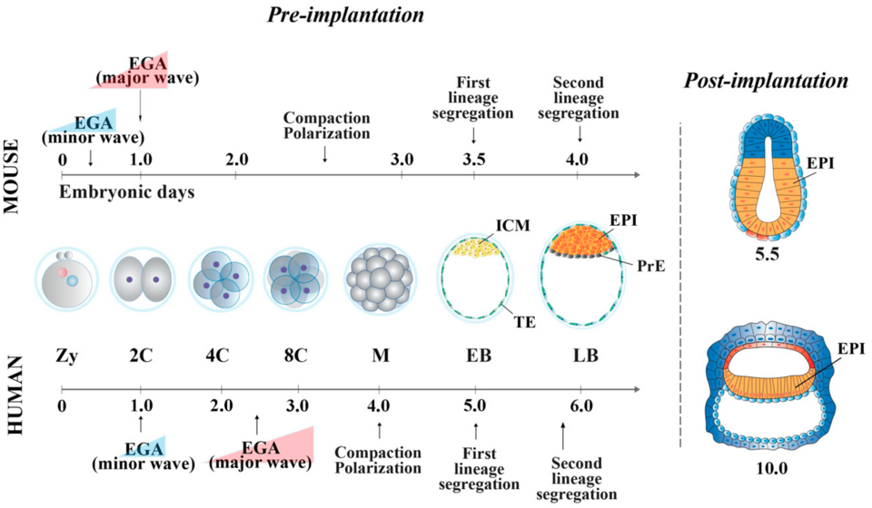 Label zygote, morula, blastocyst, and implantation. (Image: Rebuzzini, P.; Zuccotti, M.; Garagna, S., CC BY 4.0, via Wikimedia Commons)
4. Hormonal Regulation
| Hormone | Source | Function |
|---|---|---|
| FSH | Pituitary | Stimulates follicle growth (female) / spermatogenesis (male) |
| LH | Pituitary | Triggers ovulation (female) / testosterone production (male) |
| Estrogen | Ovaries | Thickens endometrium, secondary sexual traits |
| Progesterone | Corpus luteum | Maintains endometrium for pregnancy |
| Testosterone | Testes (Leydig cells) | Sperm production, muscle growth, libido |
| Oxytocin | Posterior pituitary | Stimulates uterine contractions (childbirth) and milk ejection |
| Prolactin | Pituitary | Milk production after childbirth |
5. Reproductive Disorders
A. Sexually Transmitted Diseases (STDs)
| Disease | Cause | Symptoms | Prevention |
|---|---|---|---|
| HIV/AIDS | HIV virus | Weakened immunity, weight loss | Safe sex, antiretrovirals |
| Syphilis | Treponema pallidum | Chancres, rash, neurological damage | Condoms, early treatment |
| Gonorrhea | Neisseria gonorrhoeae | Painful urination, discharge | Condoms, antibiotics |
| Chlamydia | Chlamydia trachomatis | Silent often, pelvic inflammatory disease | Condoms, screening |
B. Infertility Causes
- Male: Low sperm count, blocked vas deferens, hormonal imbalances.
- Female: Blocked fallopian tubes, PCOS (Polycystic Ovary Syndrome), endometriosis.
- Solutions: IVF, IUI, hormone therapy.
6. Assisted Reproductive Technologies (ART)
| Method | Process | Success Rate | Ethical Issues |
|---|---|---|---|
| IVF | Egg + sperm fertilized in lab; embryo implanted | ~30–40% per cycle | Embryo disposal, genetic screening |
| IUI | Sperm inserted into uterus via catheter | ~10–20% | Low cost, minimal invasiveness |
| Surrogacy | Another woman carries the pregnancy | Varies | Legal and emotional complexities |
Exam Tip
- Diagrams are crucial: Label male/female reproductive organs, menstrual cycle phases, and fertilization steps accurately.
- Hormone roles: Memorize FSH, LH, estrogen, progesterone functions and their sources.
- Comparisons: Know differences between spermatogenesis vs. oogenesis and male vs. female reproductive systems.
- NEB-style questions:
- "Describe the role of progesterone in the menstrual cycle." → Focus on endometrium maintenance.
- "How does the contraceptive pill prevent pregnancy?" → Inhibits FSH/LH → no ovulation.
- "Draw and label the stages of early embryonic development." → Zygote → morula → blastocyst.
- Common mistakes:
- Confusing primary/secondary oocytes with polar bodies.
- Forgetting testosterone’s role in males (not just sperm production).
- Mixing up fallopian tube vs. uterus functions.
Practice Questions (NEB Style)
Short Answer
- Define spermatogenesis and list the hormones involved.
- What is the role of the corpus luteum in the menstrual cycle?
- Differentiate between ovulation and menstruation.
Long Answer
- Explain the process of fertilization with a labeled diagram. What prevents polyspermy?
- Describe the menstrual cycle, highlighting the hormonal changes at each phase.
- What are STDs? Give two examples and their prevention methods.
Diagram-Based
- Draw and label the male reproductive system, showing the path of sperm from production to ejaculation.
- Sketch the stages of early embryonic development (zygote to blastocyst).
Key Formulas/Terms
- Menstrual Cycle Duration: ~28 days (varies).
- Sperm Count: 50–150 million/mL (normal).
- Fertile Window: Days 10–17 (ovulation + sperm viability).
- Acrosome Reaction: Enzymes (hyaluronidase, acrosin) digest zona pellucida.
Based on the NEB +2 Science syllabus for Biology (Bio), unit 15.
Discussion
Loading…