BiologyUnit 1610 min read
Human Respiratory System: Organs, Gas Exchange & Disorders
Unit 16 of Biology explains the human respiratory system’s anatomy, mechanics of breathing, gas transport in blood, and common diseases like asthma and tuberculosis, with NEB-style questions and solved examples.
TAKEAWAYS:
- The respiratory system includes the nose, pharynx, larynx, trachea, bronchi, bronchioles, and alveoli, where gas exchange occurs.
- Breathing involves inhalation (diaphragm contraction) and exhalation (diaphragm relaxation), regulated by the medulla oblongata.
- Oxygen binds to hemoglobin in red blood cells, while carbon dioxide is transported as bicarbonate ions.
- Disorders like asthma, tuberculosis, and emphysema disrupt normal respiration and require medical intervention.
- The respiratory system works closely with the circulatory system to supply oxygen to cells and remove carbon dioxide.
- NEB exams test definitions, functions, and comparisons (e.g., breathing vs. respiration, alveoli vs. bronchioles).
---

### **1. Introduction to the Respiratory System**
The **respiratory system** is responsible for **gas exchange**—taking in **oxygen (O₂)** and removing **carbon dioxide (CO₂)** from the body. It works with the **circulatory system** to supply oxygen to all cells and remove waste gases.

#### **Key Functions:**
- **Breathing (ventilation):** Movement of air in and out of the lungs.
- **Gas exchange:** Oxygen enters the blood, and carbon dioxide leaves it.
- **Speech and smell:** The larynx helps produce sound, and the nose detects odors.
- **pH regulation:** Helps maintain the body’s acid-base balance by controlling CO₂ levels.

---
```figure
{"type":"layers","layers":["Nose","Pharynx","Larynx","Trachea","Bronchi","Bronchioles","Alveoli"],"right":["Air enters","Air passes","Voice box","Windpipe","Branches into lungs","Smaller tubes","Gas exchange sites"],"highlight":["Alveoli"],"caption":"Pathway of air in the respiratory system"}
2. Anatomy of the Respiratory System
The respiratory system is divided into two main parts:
- Upper respiratory tract (nose, pharynx, larynx)
- Lower respiratory tract (trachea, bronchi, bronchioles, alveoli)
A. Upper Respiratory Tract
Nose:
- Functions: Filters dust, warms, and moistens air.
- Structures:
- Nasal cavity: Lined with cilia (tiny hair-like structures) and mucus to trap particles.
- Nasal septum: Divides the nose into two halves.
- Sinuses: Lighten the skull and help with voice resonance.
Pharynx (Throat):
- Common passage for air (respiratory system) and food (digestive system).
- Divided into:
- Nasopharynx (behind the nose)
- Oropharynx (behind the mouth)
- Laryngopharynx (leads to the larynx and esophagus).
Larynx (Voice Box):
- Contains vocal cords, which vibrate to produce sound.
- Epiglottis: A flap that prevents food from entering the trachea during swallowing.
flowchart TD
    A["Nose"] --> B["Pharynx"]
    B --> C["Larynx"]
    C --> D["Trachea"]
    D --> E["Bronchi"]
    E --> F["Bronchioles"]
    F --> G["Alveoli"]
    G -->|"Gas Exchange"| H["Blood"]
    H -->|"Oxygen to Cells"| I["Body"]
    I -->|"CO₂ Waste"| H
B. Lower Respiratory Tract
Trachea (Windpipe):
- A rigid tube supported by C-shaped cartilage rings to stay open.
- Lined with cilia and mucus to trap dust and pathogens.
- Divides into left and right bronchi (one for each lung).
Bronchi and Bronchioles:
- Bronchi: Two main branches from the trachea.
- Bronchioles: Smaller tubes branching from bronchi, ending in alveoli.
Alveoli (Air Sacs):
- Tiny, balloon-like structures where gas exchange occurs.
- Surrounded by capillaries (tiny blood vessels).
- Structure: Thin walls (1 cell thick) for efficient diffusion.
- Function: Oxygen diffuses into the blood, and CO₂ diffuses out.
3. Mechanics of Breathing (Ventilation)
Breathing is controlled by muscles and the nervous system.
A. Inhalation (Breathing In)
- Diaphragm contracts (moves downward).
- External intercostal muscles contract, lifting the ribs.
- Thoracic cavity expands, reducing pressure inside the lungs.
- Air rushes in through the nose/mouth to equalize pressure.
B. Exhalation (Breathing Out)
- Diaphragm relaxes (moves upward).
- External intercostal muscles relax, ribs move down.
- Thoracic cavity shrinks, increasing pressure inside the lungs.
- Air is forced out through the respiratory tract.
4. Gas Exchange in the Lungs
Gas exchange occurs in the alveoli due to diffusion.
A. Oxygen Transport
- Oxygen (O₂) diffuses from alveoli into capillary blood.
- Hemoglobin in red blood cells binds with O₂ to form oxyhemoglobin (HbO₂).
- Oxygenated blood is carried to the left atrium of the heart and pumped to the body.
B. Carbon Dioxide Transport
- Carbon dioxide (CO₂) diffuses from blood into alveoli.
- Most CO₂ is converted to bicarbonate ions (HCO₃⁻) in red blood cells.
- CO₂ is exhaled out of the body.
5. Control of Breathing
Breathing is regulated by the medulla oblongata (part of the brainstem).
A. Respiratory Center
- Monitors CO₂ levels in the blood.
- If CO₂ increases, it stimulates inhalation.
- If CO₂ decreases, it slows down breathing.
B. Chemoreceptors
- Located in the aorta and carotid arteries.
- Detect changes in pH (acidity) due to CO₂.
- Send signals to the medulla to adjust breathing rate.
flowchart LR
    A["High CO₂ in Blood"] --> B["Detected by Chemoreceptors"]
    B --> C["Signal to Medulla"]
    C --> D["Increase Breathing Rate"]
    D --> E["More CO₂ Expelled"]
    E --> A
6. Disorders of the Respiratory System
Common respiratory disorders include:
| Disease | Cause | Effects | Treatment |
|---|---|---|---|
| Asthma | Allergens, pollution, stress | Bronchial spasms, wheezing, difficulty breathing | Inhalers, avoiding triggers |
| Tuberculosis (TB) | Mycobacterium tuberculosis bacteria | Coughing, fever, weight loss, lung damage | Antibiotics, rest, nutrition |
| Emphysema | Smoking, air pollution | Destruction of alveoli, shortness of breath | Oxygen therapy, lung transplant |
| Pneumonia | Bacterial/viral infection | Fluid in alveoli, difficulty breathing | Antibiotics, rest, hydration |
| Bronchitis | Viral/bacterial infection | Inflammation of bronchi, coughing | Rest, fluids, medication |
7. Practical Applications
A. Respiratory Volume Measurements
- Tidal Volume (TV): Air inhaled/exhaled in normal breathing (~500 mL).
- Inspiratory Reserve Volume (IRV): Extra air inhaled after normal breath (~3000 mL).
- Expiratory Reserve Volume (ERV): Extra air exhaled after normal breath (~1200 mL).
- Residual Volume (RV): Air remaining in lungs after forced exhalation (~1200 mL).
Example Calculation: If a person’s Total Lung Capacity (TLC) is 6000 mL, and their Vital Capacity (VC = TV + IRV + ERV) is 4800 mL, what is their Residual Volume (RV)? Solution:
B. Effect of Exercise on Breathing
- Increased breathing rate to supply more oxygen.
- Deeper inhalations to maximize air intake.
- More efficient gas exchange due to increased blood flow.
8. NEB-Style Questions & Solutions
Short Answer Questions
Define the following terms:
- Alveoli: Tiny air sacs in the lungs where gas exchange occurs.
- Diaphragm: A muscle that contracts and relaxes to help with breathing.
- Hemoglobin: A protein in red blood cells that binds with oxygen.
Differentiate between:
Breathing Respiration Physical process of air movement Chemical process of gas exchange Involves lungs and muscles Occurs in cells (mitochondria) Does not require energy Requires energy (ATP) Why is the trachea supported by cartilage rings?
- To keep it open so air can flow freely.
- Prevents collapse during breathing.
Long Answer Questions
Describe the process of gas exchange in the alveoli.
- Oxygen from inhaled air diffuses through alveolar membrane into capillary blood.
- Carbon dioxide from blood diffuses into alveoli to be exhaled.
- Hemoglobin in red blood cells binds with oxygen for transport.
Explain how the respiratory system works with the circulatory system.
- Lungs oxygenate blood and remove CO₂.
- Heart pumps oxygenated blood to body cells and returns deoxygenated blood to lungs.
- Capillaries facilitate gas exchange between blood and tissues.
Diagram-Based Questions
Label the following parts of the respiratory system:
- Nose, Pharynx, Larynx, Trachea, Bronchi, Bronchioles, Alveoli, Diaphragm. (Use the diagram from Section 2.)
Draw and explain the mechanism of inhalation and exhalation. (Use the Mermaid flowchart from Section 3.)
Exam Tip
✅ For NEB exams:
- Memorize key terms (alveoli, diaphragm, hemoglobin, tidal volume).
- Understand the difference between breathing (ventilation) and respiration (gas exchange).
- Practice labeling diagrams of the respiratory system.
- Explain gas exchange and breathing mechanics step-by-step.
- Compare respiratory disorders (causes, effects, treatments).
- Solve numerical problems on lung volumes (TLC, VC, RV).
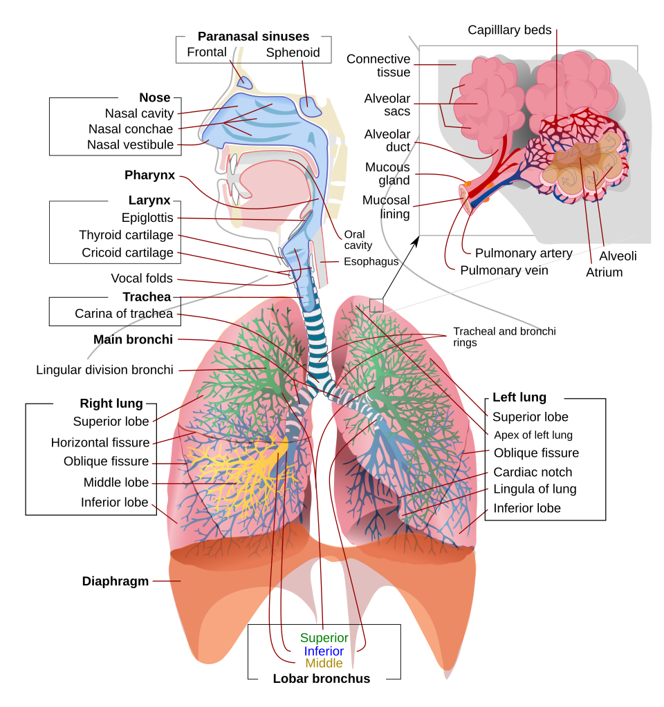 Label the parts of the respiratory tract. (Image: LadyofHats, Jmarchn, Public domain, via Wikimedia Commons)
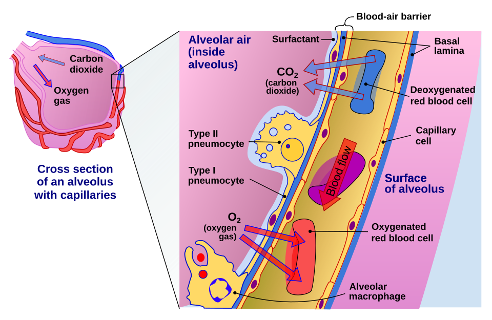 Show how oxygen and CO₂ move between alveoli and blood. (Image: Delmalani18, CC BY-SA 4.0, via Wikimedia Commons)
Based on the NEB +2 Science syllabus for Biology (Bio), unit 16.
Discussion
Loading…