BiologyUnit 1114 min read
Human Circulatory System: Heart, Blood, Vessels & Disorders
Unit 11 of Biology covers the human circulatory system, including the heart’s anatomy and function, blood composition, blood vessel types, blood circulation pathways, and common circulatory disorders—essential for NEB exams.
```mermaid
mindmap
  root((Human Circulatory System))
    Heart
      Structure
      Function
    Blood
      Composition
      Functions
    Blood Vessels
      Arteries
      Veins
      Capillaries
    Circulation
      Pulmonary
      Systemic
    Disorders
      Hypertension
      Atherosclerosis
      Anaemia
The Human Circulatory System: An Overview
The circulatory system (also called the cardiovascular system) is a closed transport system that carries blood, nutrients, gases, and waste throughout the body. It consists of:
- Heart (the pump)
- Blood (the transport medium)
- Blood vessels (the pathways: arteries, veins, capillaries)
This system works 24/7 to keep us alive by delivering oxygen and food to cells and removing carbon dioxide and waste.
1. The Heart: The Powerhouse of Circulation
The heart is a muscular pump the size of a fist, located in the thoracic cavity between the lungs. It beats ~72 times per minute at rest, pumping ~5–6 litres of blood per minute.
A. Structure of the Heart
graph TD
  A["Heart"] --> B["Pericardium"]
  A --> C["Myocardium"]
  A --> D["Endocardium"]
  B --> E["Fibrous Pericardium"]
  B --> F["Serous Pericardium"]
  F --> G["Parietal Layer"]
  F --> H["Visceral Layer (Epicardium)"]
  C --> I["Cardiac Muscle"]
  D --> J["Inner Lining"]
  A --> K["4 Chambers"]
  K --> L["Atria: Right & Left"]
  K --> M["Ventricles: Right & Left"]
  L --> N["Right Atrium"]
  L --> O["Left Atrium"]
  M --> P["Right Ventricle"]
  M --> Q["Left Ventricle"]
  A --> R["Valves"]
  R --> S["Atrioventricular Valves: Tricuspid & Bicuspid"]
  R --> T["Semilunar Valves: Pulmonary & Aortic"]
Key Parts of the Heart
| Part | Function |
|---|---|
| Pericardium | Protective double-layered sac around the heart. |
| Myocardium | Thick muscular layer that contracts to pump blood. |
| Endocardium | Smooth inner lining that prevents blood clotting. |
| Atria (2) | Receive blood (right atrium = deoxygenated; left atrium = oxygenated). |
| Ventricles (2) | Pump blood (right ventricle → lungs; left ventricle → body). |
| Valves | Prevent backflow of blood (e.g., tricuspid, bicuspid, pulmonary, aortic). |
Heart Valves and Their Functions
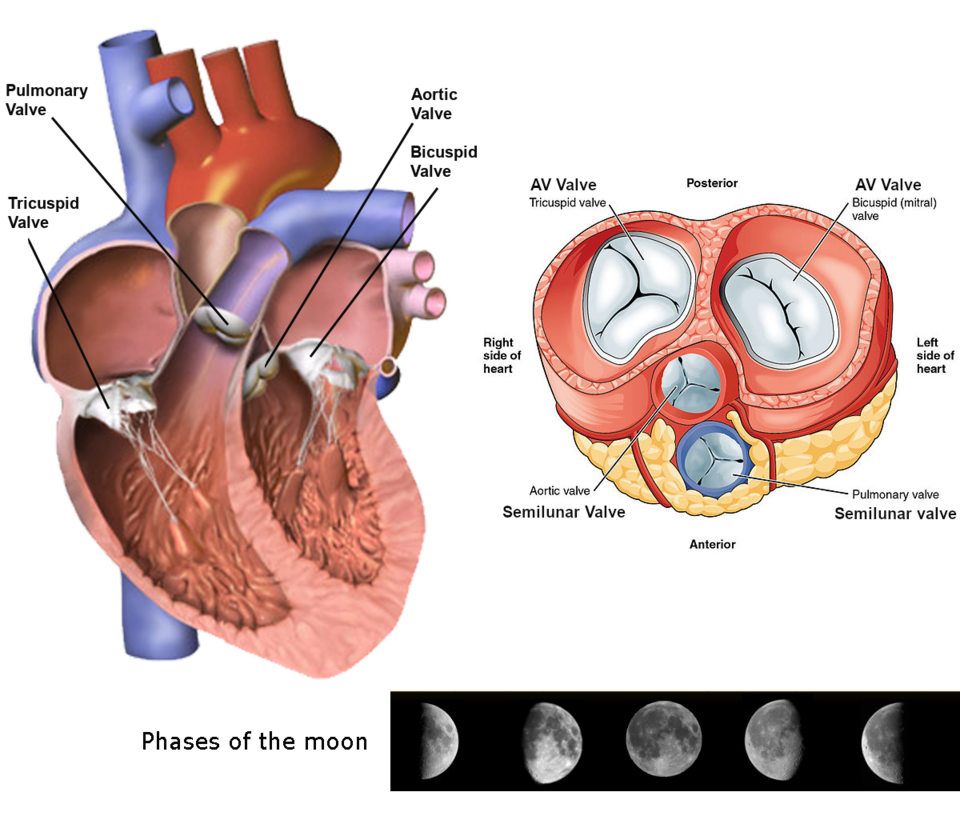 The four chambers and valves of the human heart. (Image: RWhitwam, CC BY-SA 4.0, via Wikimedia Commons)
B. Cardiac Cycle: How the Heart Works
The heart goes through a continuous cycle of contraction (systole) and relaxation (diastole).
Steps of the Cardiac Cycle
Atrial Systole (Atria contract)
- Blood is pushed into the ventricles.
- AV valves (tricuspid & bicuspid) open; semilunar valves closed.
Ventricular Systole (Ventricles contract)
- Blood is pumped out:
- Right ventricle → Pulmonary artery → Lungs (deoxygenated blood).
- Left ventricle → Aorta → Body (oxygenated blood).
- Semilunar valves open; AV valves close (prevents backflow).
- Blood is pumped out:
Diastole (Heart relaxes)
- All chambers relax and fill with blood.
- AV valves open; semilunar valves closed.
graph LR
  A["Atrial Systole"] -->|"Blood enters ventricles"| B["Ventricular Systole"]
  B -->|"Blood pumped out"| C["Diastole"]
  C -->|"Heart fills"| A
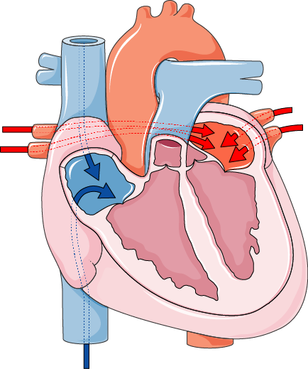 The phases of the cardiac cycle showing valve movements. (Image: Laboratoires Servier, CC BY-SA 3.0, via Wikimedia Commons)
C. Heart Sounds and ECG
- Lubb (First sound): Caused by closure of AV valves (start of ventricular systole).
- Dubb (Second sound): Caused by closure of semilunar valves (end of ventricular systole).
Electrocardiogram (ECG/EKG) records the electrical activity of the heart:
- P-wave: Atrial depolarisation (contraction).
- QRS complex: Ventricular depolarisation (contraction).
- T-wave: Ventricular repolarisation (relaxation).
2. Blood: The Body’s Transport Fluid
Blood is a connective tissue made of plasma (55%) and formed elements (45%).
A. Composition of Blood
| Component | Percentage | Function |
|---|---|---|
| Plasma | 55% | Transport of nutrients, hormones, waste, and heat. |
| Red Blood Cells (RBCs) | 45% | Carry oxygen (haemoglobin) and CO₂. |
| White Blood Cells (WBCs) | <1% | Fight infections (phagocytosis, antibodies). |
| Platelets | <1% | Help in blood clotting (prevent excessive bleeding). |
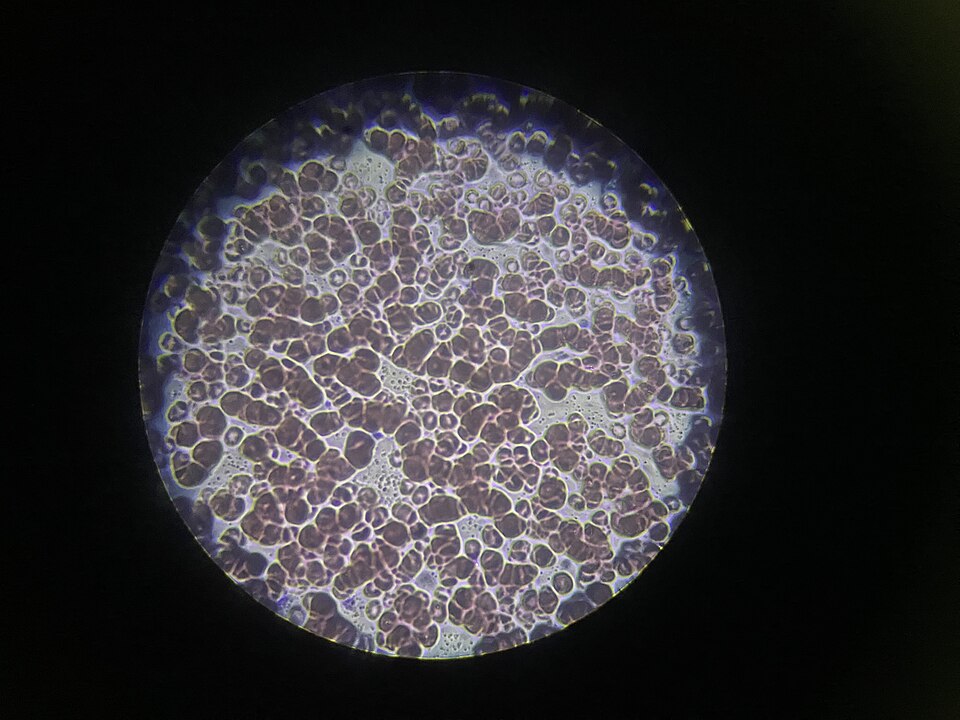 A microscopic view showing RBCs, WBCs, and platelets. (Image: Rockiesss2071, CC BY 4.0, via Wikimedia Commons)
B. Functions of Blood
Transportation
- Oxygen (from lungs to tissues via haemoglobin).
- Nutrients (glucose, amino acids from digestion).
- Waste (CO₂ to lungs, urea to kidneys).
- Hormones (e.g., insulin, adrenaline).
Protection
- WBCs fight pathogens.
- Antibodies neutralise toxins.
- Platelets prevent bleeding.
Regulation
- Maintains pH, temperature, and water balance.
C. Blood Groups and Transfusion
Blood groups are classified based on antigens (A, B, AB, O) and Rh factor (+/-).
| Blood Group | Antigens | Antibodies | Can Donate To | Can Receive From |
|---|---|---|---|---|
| A | A | Anti-B | A, AB | A, O |
| B | B | Anti-A | B, AB | B, O |
| AB | A & B | None | AB | A, B, AB, O |
| O | None | Anti-A & Anti-B | A, B, AB, O | O |
Universal Donor: O- (no antigens). Universal Recipient: AB+ (no antibodies).
3. Blood Vessels: The Highways of Circulation
Blood vessels form a closed loop that carries blood to and from the heart.
A. Types of Blood Vessels
| Type | Structure | Function | Blood Type |
|---|---|---|---|
| Arteries | Thick, elastic walls | Carry blood away from heart (high pressure). | Oxygenated (except pulmonary artery) |
| Veins | Thin walls, valves | Carry blood towards heart (low pressure). | Deoxygenated (except pulmonary vein) |
| Capillaries | Microscopic, one-cell thick | Exchange of gases, nutrients, waste. | Both oxygenated & deoxygenated |
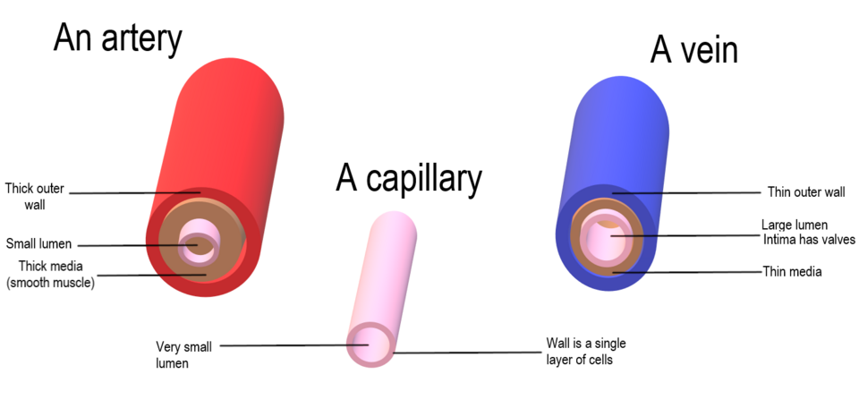 Comparing the structure of blood vessels. (Image: Christinelmiller, CC BY-SA 4.0, via Wikimedia Commons)
B. Blood Pressure and Pulse
Blood Pressure (BP): Force exerted by blood on vessel walls.
- Systolic Pressure: Pressure during ventricular contraction (e.g., 120 mmHg).
- Diastolic Pressure: Pressure during relaxation (e.g., 80 mmHg).
- Normal BP: 120/80 mmHg.
Pulse: The rhythmic expansion of arteries due to heartbeats (felt in wrist, neck).
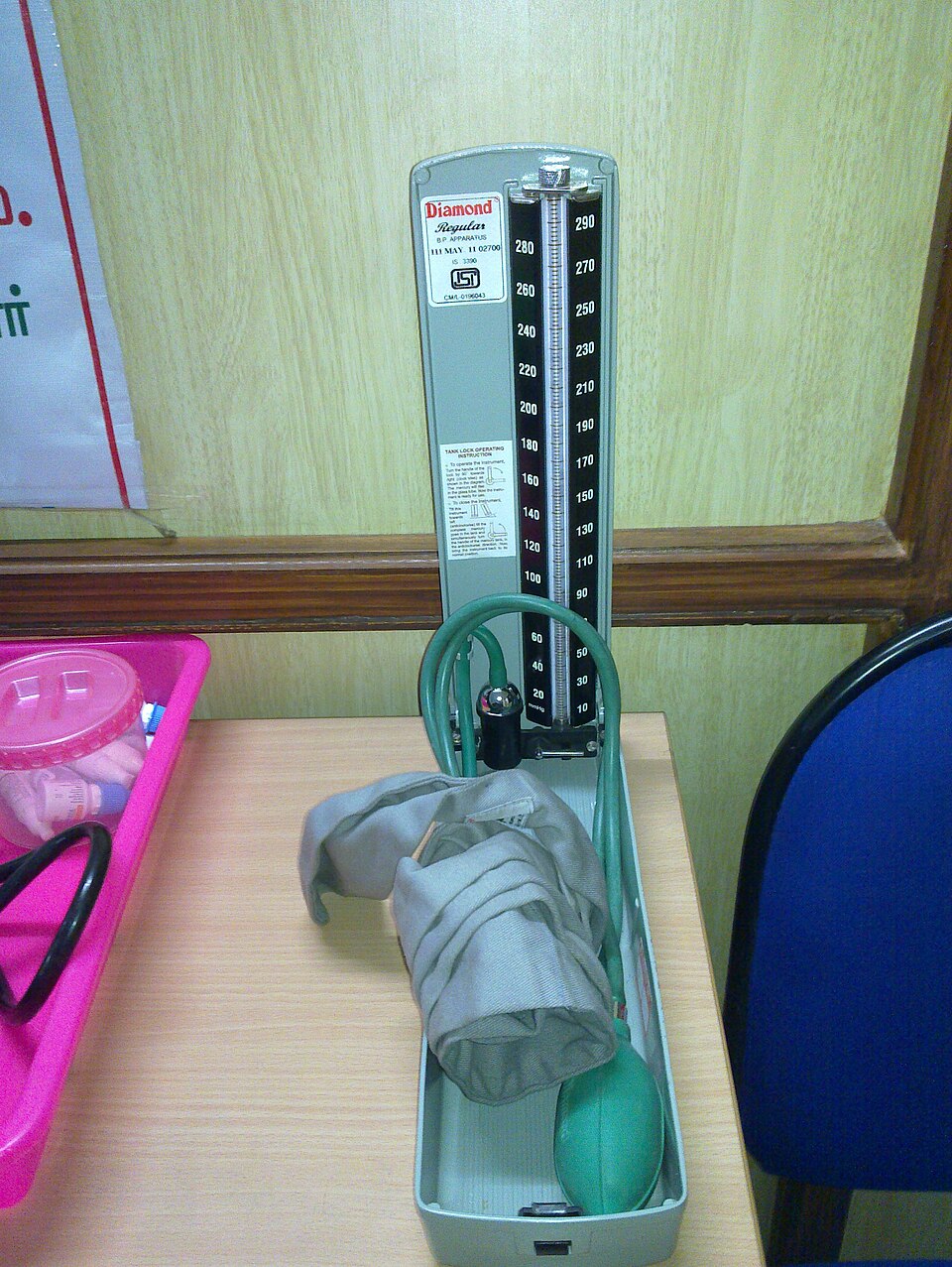 A blood pressure monitor measuring systolic and diastolic pressure. (Image: Parvathisri, CC BY-SA 3.0, via Wikimedia Commons)
4. Circulation Pathways
There are two main circuits:
Pulmonary Circulation (Heart → Lungs → Heart)
- Right ventricle → Pulmonary artery → Lungs (gas exchange) → Pulmonary vein → Left atrium.
- Function: Oxygenates blood and removes CO₂.
Systemic Circulation (Heart → Body → Heart)
- Left ventricle → Aorta → Body tissues → Vena cava → Right atrium.
- Function: Delivers O₂ and nutrients; removes CO₂ and waste.
graph TD
  A["Right Atrium"] -->|"Deoxygenated blood"| B["Right Ventricle"]
  B -->|"Pulmonary Artery"| C["Lungs"]
  C -->|"Oxygenated blood"| D["Left Atrium"]
  D -->|"Left Ventricle"| E["Aorta"]
  E -->|"To Body"| F["Body Tissues"]
  F -->|"Deoxygenated blood"| G["Vena Cava"]
  G --> A
5. Common Circulatory Disorders
| Disorder | Cause | Effects | Prevention/Treatment |
|---|---|---|---|
| Hypertension | High BP due to stress, obesity, genetics. | Damages arteries, heart failure, stroke. | Low salt, exercise, medication. |
| Atherosclerosis | Plaque buildup in arteries. | Reduces blood flow, heart attack, stroke. | Healthy diet, no smoking. |
| Anaemia | Low RBCs or haemoglobin. | Fatigue, weakness, pale skin. | Iron-rich diet, supplements. |
| Coronary Artery Disease | Blocked coronary arteries. | Chest pain (angina), heart attack. | Stents, bypass surgery. |
| Varicose Veins | Weak vein walls (valve failure). | Swollen, twisted veins. | Compression stockings, exercise. |
Exam Tip: How to Score Full Marks
✅ Diagrams are mandatory – Always draw and label:
- Heart structure (chambers, valves).
- Blood circulation pathways.
- ECG waveform.
✅ Compare and contrast – Questions often ask:
- Arteries vs. Veins (structure, function, blood type).
- Pulmonary vs. Systemic Circulation (pathways, purpose).
✅ Define key terms – Expect questions on:
- Cardiac cycle, blood pressure, haemoglobin, blood groups.
✅ Apply concepts – Example:
- "Why does a person with blood group AB not need antibodies?"
- "How does atherosclerosis affect blood flow?"
✅ Practical questions – Be ready for:
- ECG interpretation (identify P, QRS, T waves).
- Blood pressure calculations (systolic vs. diastolic).
- Transfusion compatibility (matching donor-recipient groups).
NEB-Style Questions (Practice)
Short Answer (5 marks each)
- Draw a labelled diagram of the human heart and explain the function of the semilunar valves.
- What is the difference between arteries and veins? Why are veins equipped with valves?
- Explain the cardiac cycle with reference to the AV valves and semilunar valves.
- How does anaemia affect the efficiency of the circulatory system?
Long Answer (10 marks)
- Describe the composition and functions of blood. Why is blood considered a connective tissue?
- Explain the double circulation in humans. How does it ensure efficient oxygen supply to tissues?
- What are blood groups? Why is O- called the universal donor? Can a person with AB+ blood receive blood from all groups? Justify.
Worked Example: Blood Pressure Calculation
Question: If a person’s systolic pressure is 140 mmHg and diastolic pressure is 90 mmHg, what is their blood pressure reading, and what does it indicate?
Solution:
- Blood pressure is written as systolic/diastolic.
- 140/90 mmHg is classified as Stage 1 Hypertension (high BP, risk of heart disease).
- Prevention: Reduce salt intake, exercise, manage stress.
Final Summary Table
| Topic | Key Points |
|---|---|
| Heart Structure | 4 chambers, 4 valves, pericardium, myocardium. |
| Cardiac Cycle | Atrial systole → Ventricular systole → Diastole. |
| Blood Composition | Plasma (55%), RBCs (45%), WBCs, platelets. |
| Blood Vessels | Arteries (high pressure), veins (valves), capillaries (exchange). |
| Circulation | Pulmonary (heart-lungs), Systemic (heart-body). |
| Disorders | Hypertension, atherosclerosis, anaemia, varicose veins. |
Remember: The circulatory system is like a highway network—the heart is the traffic controller, blood is the vehicles, and blood vessels are the roads. Keep this analogy in mind for exams! 🚗💉🩸
Based on the NEB +2 Science syllabus for Biology (Bio), unit 11.
Discussion
Loading…