BiologyUnit 1315 min read
Nervous System & Sense Organs: Structure, Function & Coordination
Unit 13 of Biology explores how the human nervous system works—from neurons to reflexes—and how our five senses (vision, hearing, taste, smell, touch) detect and interpret the world, with clear diagrams, real examples, and NEB-style questions.
TAKEAWAYS:
- The nervous system is divided into central (brain + spinal cord) and peripheral (somatic + autonomic) parts, each with distinct functions.
- Neurons transmit signals via electrical impulses (action potentials) and chemical neurotransmitters (e.g., acetylcholine).
- Reflex arcs are rapid, automatic responses (e.g., knee-jerk) that bypass the brain for faster protection.
- Sense organs (eyes, ears, nose, tongue, skin) convert physical stimuli into nerve impulses using specialized receptors.
- Hormones and nerves both coordinate body functions, but nerves act faster (milliseconds) while hormones take seconds to hours.
- Common disorders (e.g., myopia, glaucoma, deafness) can be prevented or managed with lifestyle changes and early detection.
1. The Nervous System: Structure and Function
The nervous system is the control and communication system of the body. It detects changes inside and outside the body, processes this information, and responds accordingly.
A. Divisions of the Nervous System
The nervous system is divided into two main parts:
graph TD
    A["Nervous System"] --> B["Central Nervous System (CNS)"]
    A --> C["Peripheral Nervous System (PNS)"]
    B --> D["Brain"]
    B --> E["Spinal Cord"]
    C --> F["Somatic Nervous System (Voluntary)"]
    C --> G["Autonomic Nervous System (Involuntary)"]
    G --> H["Sympathetic (Fight or Flight)"]
    G --> I["Parasympathetic (Rest and Digest)"]
Central Nervous System (CNS):
- Consists of the brain and spinal cord.
- Acts as the control center for the body.
- Processes information and sends out commands.
Peripheral Nervous System (PNS):
- Connects the CNS to the rest of the body.
- Divided into:
- Somatic Nervous System (SNS): Controls voluntary movements (e.g., walking, writing).
- Autonomic Nervous System (ANS): Controls involuntary functions (e.g., heartbeat, digestion).
- Sympathetic NS: Prepares the body for "fight or flight" (e.g., increased heart rate, dilated pupils).
- Parasympathetic NS: Promotes "rest and digest" (e.g., slowed heart rate, digestion).
B. Neurons: The Basic Units of the Nervous System
Neurons (nerve cells) are the building blocks of the nervous system. They transmit information as electrical impulses (action potentials) and use chemical signals (neurotransmitters) to communicate.
graph TD
    A["Neuron Structure"] --> B["Dendrites"]
    A --> C["Cell Body (Soma)"]
    A --> D["Nucleus"]
    A --> E["Axon"]
    A --> F["Myelin Sheath"]
    A --> G["Axon Terminals"]
A --> H["Synapse"]- Dendrites: Receive signals from other neurons.
- Cell Body (Soma): Contains the nucleus and keeps the neuron alive.
- Axon: Long fiber that carries the electrical impulse away from the cell body.
- Myelin Sheath: Fatty layer that insulates the axon and speeds up signal transmission.
- Axon Terminals: Release neurotransmitters (e.g., acetylcholine, dopamine) into the synapse.
- Synapse: The gap between neurons where chemical signals are transmitted.
How a Neuron Works:
- A stimulus (e.g., touch, light) causes a change in membrane potential.
- If the stimulus is strong enough, an action potential (electrical impulse) is generated.
- The impulse travels down the axon to the axon terminals.
- Neurotransmitters are released into the synapse and bind to receptors on the next neuron.
- The signal is passed on, creating a nerve impulse.
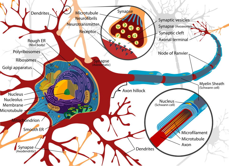 Labelled structure of a neuron showing dendrites, cell body, axon, myelin sheath, and axon terminals. (Image: LadyofHats, Public domain, via Wikimedia Commons)
C. Resting and Action Potential
- Resting Potential: The negative charge inside a neuron at rest (~ -70 mV).
- Action Potential: A rapid change in membrane potential that travels along the axon (~ +30 mV).
graph TD
    A["Resting Potential (-70 mV)"] -->|"Stimulus"| B["Depolarization (Na+ enters)"]
    B --> C["Action Potential (+30 mV)"]
    C --> D["Repolarization (K+ exits)"]
    D --> E["Resting Potential (-70 mV)"]
Example: When you touch a hot object, pain receptors in your skin send a signal to your spinal cord. If the stimulus is strong enough, an action potential is generated, and you quickly pull your hand away.
2. Reflex Actions
Reflexes are rapid, automatic responses to stimuli that do not involve conscious thought. They protect the body from harm.
A. Reflex Arc
A reflex arc is the neural pathway for a reflex action. It involves:
- Receptor: Detects the stimulus (e.g., pain in the finger).
- Sensory Neuron: Carries the signal to the spinal cord.
- Interneuron (in spinal cord): Processes the signal.
- Motor Neuron: Sends the signal to a muscle or gland.
- Effector: Responds to the stimulus (e.g., muscle contracts to pull the hand away).
graph TD
    A["Stimulus (e.g., Pain)"] --> B["Receptor"]
    B --> C["Sensory Neuron"]
    C --> D["Spinal Cord (Interneuron)"]
    D --> E["Motor Neuron"]
    E --> F["Effector (Muscle)"]
    F --> G["Response (Pull Hand Away)"]
Example: Knee-Jerk Reflex
- A doctor taps your knee with a hammer.
- The stretch receptor in your quadriceps muscle sends a signal to the spinal cord.
- The spinal cord sends a signal back to the muscle, causing it to contract.
- Your leg kicks forward without you thinking about it.
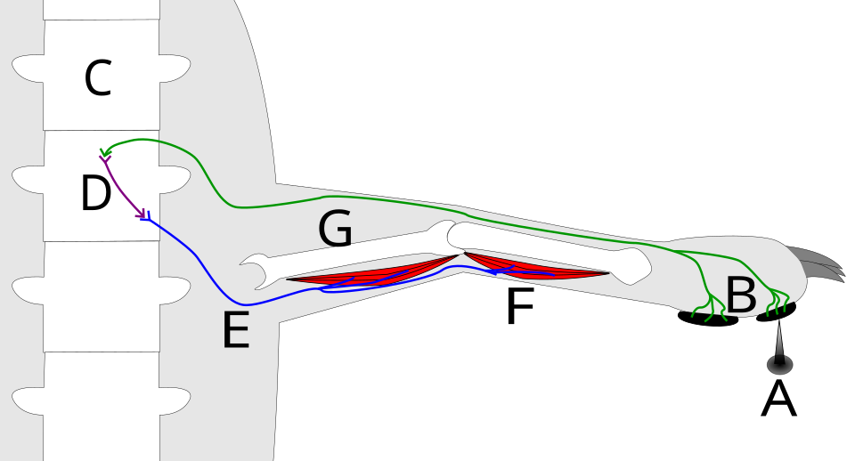 Labelled diagram of a knee-jerk reflex showing receptors, sensory neuron, spinal cord, motor neuron, and effector. (Image: Verona Dethran, CC BY-SA 4.0, via Wikimedia Commons)
B. Importance of Reflexes
- Protection: Prevents injury (e.g., pulling hand away from heat).
- Speed: Faster than conscious responses (no brain involvement).
- Automatic: Do not require learning (innate).
3. The Brain: Structure and Functions
The brain is the command center of the nervous system. It is divided into three main parts:
graph TD
    A["Brain"] --> B["Forebrain"]
    A --> C["Midbrain"]
    A --> D["Hindbrain"]
    B --> E["Cerebrum"]
    B --> F["Thalamus"]
    B --> G["Hypothalamus"]
    D --> H["Cerebellum"]
    D --> I["Medulla Oblongata"]
    D --> J["Pons"]
A. Forebrain
Cerebrum:
- Largest part of the brain.
- Responsible for thought, memory, reasoning, and voluntary movements.
- Divided into four lobes:
- Frontal Lobe: Decision-making, problem-solving.
- Parietal Lobe: Touch, temperature, body position.
- Temporal Lobe: Hearing, memory.
- Occipital Lobe: Vision.
Thalamus:
- Acts as a relay station for sensory information (except smell).
Hypothalamus:
- Controls hormone release, hunger, thirst, and body temperature.
B. Midbrain
- Controls visual and auditory reflexes.
- Helps maintain posture and movement.
C. Hindbrain
Cerebellum:
- Coordinates balance, posture, and fine motor skills (e.g., playing an instrument).
Medulla Oblongata:
- Controls automatic functions (e.g., heartbeat, breathing).
Pons:
- Helps regulate sleep and breathing.
4. The Spinal Cord
- A long, thin structure that runs from the brainstem to the lower back.
- Functions:
- Transmits signals between the brain and the rest of the body.
- Contains reflex centers (e.g., knee-jerk reflex).
Structure:
- Gray Matter: Inner part (shaped like a butterfly), contains neuron cell bodies.
- White Matter: Outer part, contains myelinated axons that transmit signals.
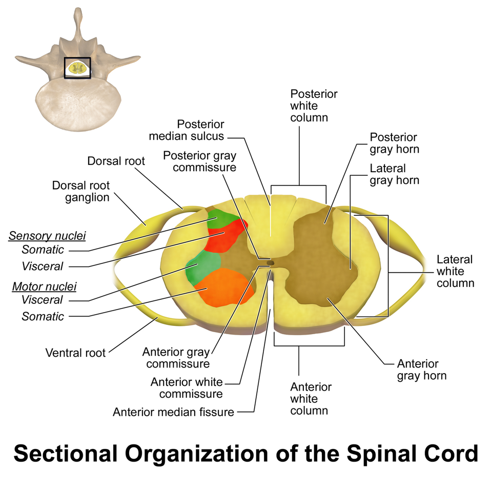 Labelled cross-section showing gray matter, white matter, and spinal nerves. (Image: BruceBlaus, CC BY-SA 4.0, via Wikimedia Commons)
5. Sense Organs
Sense organs detect stimuli (changes in the environment) and convert them into nerve impulses that the brain can interpret.
A. The Eye: Vision
The eye detects light and converts it into electrical signals that the brain interprets as images.
graph TD
    A["Light Enters"] --> B["Cornea"]
    B --> C["Pupil"]
    C --> D["Lens"]
    D --> E["Retina"]
    E --> F["Optic Nerve"]
    F --> G["Brain (Interprets Image)"]
Parts of the Eye:
- Cornea: Transparent outer layer that bends light.
- Pupil: Opening that controls how much light enters.
- Lens: Focuses light onto the retina.
- Retina: Contains photoreceptor cells (rods and cones) that detect light.
- Optic Nerve: Carries signals from the retina to the brain.
How We See:
- Light enters the eye and is focused by the lens onto the retina.
- Rods detect black and white (low light).
- Cones detect color (bright light).
- Signals are sent to the brain via the optic nerve, which interprets them as an image.
Common Eye Disorders:
| Disorder | Cause | Prevention/Treatment |
|---|---|---|
| Myopia | Elongated eyeball or strong lens | Wear glasses, avoid excessive screen time |
| Hyperopia | Shortened eyeball | Glasses, eye exercises |
| Glaucoma | Increased eye pressure | Regular eye check-ups, medication |
| Cataract | Clouding of the lens | Surgery, avoid UV exposure |
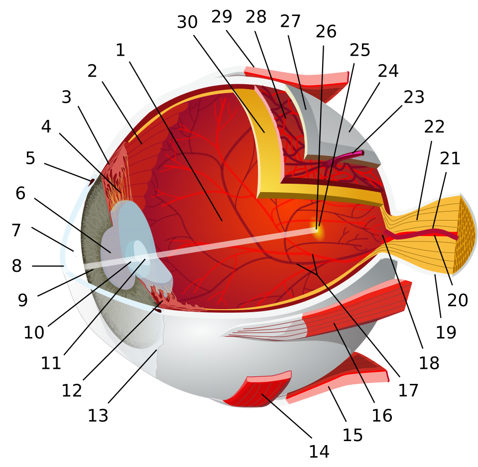 Labelled cross-section of the human eye showing cornea, lens, retina, and optic nerve. (Image: Chabacano, CC BY-SA 3.0, via Wikimedia Commons)
B. The Ear: Hearing and Balance
The ear is divided into three parts:
graph TD
    A["Ear"] --> B["Outer Ear"]
    A --> C["Middle Ear"]
    A --> D["Inner Ear"]
    B --> E["Pinna"]
    B --> F["Eardrum"]
    C --> G["Ossicles (Hammer, Anvil, Stirrup)"]
    D --> H["Cochlea"]
    D --> I["Semicircular Canals"]
Outer Ear:
- Pinna: Collects sound waves.
- Eardrum: Vibrates when sound waves hit it.
Middle Ear:
- Ossicles (Hammer, Anvil, Stirrup): Amplify vibrations and send them to the cochlea.
Inner Ear:
- Cochlea: Converts vibrations into nerve impulses (hearing).
- Semicircular Canals: Help maintain balance.
How We Hear:
- Sound waves enter the pinna and hit the eardrum.
- The ossicles amplify the vibrations.
- The cochlea converts vibrations into nerve impulses.
- The auditory nerve sends signals to the brain, which interprets them as sound.
Common Ear Disorders:
| Disorder | Cause | Prevention/Treatment |
|---|---|---|
| Deafness | Damage to cochlea or nerves | Hearing aids, cochlear implants |
| Ear Infection | Bacteria or viruses | Antibiotics, avoid swimming in dirty water |
| Tinnitus | Aging, loud noise | Avoid loud noises, use ear protection |
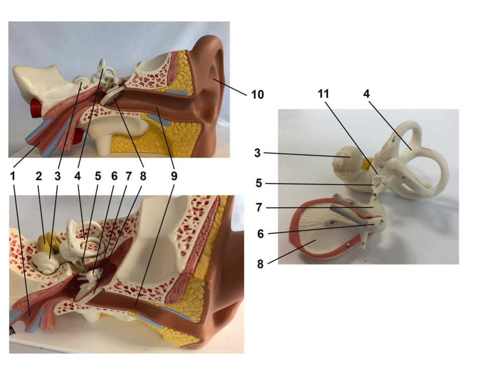 Labelled cross-section of the human ear showing outer, middle, and inner ear parts. (Image: RWhitwam, CC BY-SA 4.0, via Wikimedia Commons)
C. The Nose: Smell
- Olfactory Receptors: Detect chemicals in the air.
- Olfactory Nerve: Sends signals to the brain, which interprets them as smells.
How We Smell:
- Air enters the nose and dissolves in mucus.
- Olfactory receptors detect chemicals.
- Signals are sent to the olfactory bulb in the brain.
- The brain interprets the signals as smells.
D. The Tongue: Taste
- Taste Buds: Detect sweet, sour, salty, bitter, and umami.
- Gustatory Cells: Send signals to the brain via the gustatory nerve.
How We Taste:
- Food dissolves in saliva.
- Taste buds detect chemicals.
- Signals are sent to the brain, which interprets them as taste.
E. The Skin: Touch
- Mechanoreceptors: Detect pressure, vibration, and texture.
- Thermoreceptors: Detect heat and cold.
- Nociceptors: Detect pain.
How We Feel:
- Stimuli (e.g., touch, heat) activate receptors in the skin.
- Signals are sent to the spinal cord and then to the brain.
- The brain interprets the signals as touch, temperature, or pain.
6. Nervous System vs. Endocrine System
Both systems coordinate body functions, but they work differently:
| Feature | Nervous System | Endocrine System |
|---|---|---|
| Speed | Fast (milliseconds) | Slow (seconds to hours) |
| Signals | Electrical (action potentials) + chemical (neurotransmitters) | Chemical (hormones) in blood |
| Target | Specific organs/muscles | General (affects many cells) |
| Duration | Short-lived | Long-lasting |
| Example | Knee-jerk reflex | Adrenaline release during stress |
7. Common Disorders of the Nervous System
| Disorder | Cause | Symptoms | Treatment/Prevention |
|---|---|---|---|
| Parkinson’s Disease | Death of dopamine-producing neurons | Tremors, stiffness, slow movement | Medication, physical therapy |
| Epilepsy | Abnormal electrical activity in brain | Seizures | Medication, lifestyle changes |
| Alzheimer’s Disease | Loss of neurons in brain | Memory loss, confusion | No cure, early diagnosis helps |
| Multiple Sclerosis | Immune system attacks myelin sheath | Muscle weakness, vision problems | Medication, physical therapy |
Exam Tip: How to Score Full Marks
Diagrams are Key:
- Always draw labelled diagrams for the eye, ear, neuron, brain, and reflex arc.
- Use arrows to show the path of signals (e.g., in reflex arcs or the ear).
Compare and Contrast:
- Questions often ask to differentiate between the nervous and endocrine systems, or between rods and cones.
- Use tables (like the one above) to organize your answers.
Explain Processes Step-by-Step:
- For reflex actions, describe the path (receptor → sensory neuron → interneuron → motor neuron → effector).
- For vision, explain how light is focused and how signals reach the brain.
Common Disorders:
- Know the causes, symptoms, and treatments of at least 3 disorders (e.g., myopia, Parkinson’s, epilepsy).
- Relate them to daily life (e.g., "Wearing glasses prevents myopia").
Practical Applications:
- Explain how reflexes protect us (e.g., pulling hand away from heat).
- Discuss how sense organs work together (e.g., smell enhances taste).
NEB-Style Questions to Practice:
- Short Answer:
- "Draw and label a neuron."
- "Explain the knee-jerk reflex."
- Long Answer:
- "Describe the structure and function of the human eye. How does myopia occur?"
- "Compare the nervous and endocrine systems with examples."
- Diagram-Based:
- "Label the parts of the ear and explain how we hear."
- Short Answer:
Final Note: The nervous system is like the body’s command center, while sense organs are its sensors. Understanding how they work helps you appreciate how quickly and efficiently your body responds to the world. Practice drawing diagrams and explaining processes step-by-step to ace your NEB exam!
Based on the NEB +2 Science syllabus for Biology (Bio), unit 13.
Discussion
Loading…